Beranda
/ Diagram Of Shoulder Muscles And Tendons - Shoulder Muscles Diagrams | 101 Diagrams - Specifically, the four rotator cuff muscles include the following
Diagram Of Shoulder Muscles And Tendons - Shoulder Muscles Diagrams | 101 Diagrams - Specifically, the four rotator cuff muscles include the following
Insurance Gas/Electricity Loans Mortgage Attorney Lawyer Donate Conference Call Degree Credit Treatment Software Classes Recovery Trading Rehab Hosting Transfer Cord Blood Claim compensation mesothelioma mesothelioma attorney Houston car accident lawyer moreno valley can you sue a doctor for wrong diagnosis doctorate in security top online doctoral programs in business educational leadership doctoral programs online car accident doctor atlanta car accident doctor atlanta accident attorney rancho Cucamonga truck accident attorney san Antonio ONLINE BUSINESS DEGREE PROGRAMS ACCREDITED online accredited psychology degree masters degree in human resources online public administration masters degree online bitcoin merchant account bitcoin merchant services compare car insurance auto insurance troy mi seo explanation digital marketing degree floridaseo company fitness showrooms stamfordct how to work more efficiently seowordpress tips meaning of seo what is an seo what does an seo do what seo stands for best seotips google seo advice seo steps, The secure cloud-based platform for smart service delivery. Safelink is used by legal, professional and financial services to protect sensitive information, accelerate business processes and increase productivity. Use Safelink to collaborate securely with clients, colleagues and external parties. Safelink has a menu of workspace types with advanced features for dispute resolution, running deals and customised client portal creation. All data is encrypted (at rest and in transit and you retain your own encryption keys. Our titan security framework ensures your data is secure and you even have the option to choose your own data location from Channel Islands, London (UK), Dublin (EU), Australia.
Diagram Of Shoulder Muscles And Tendons - Shoulder Muscles Diagrams | 101 Diagrams - Specifically, the four rotator cuff muscles include the following. The goals of shoulder surgery are to reduce pain, increase function, mobility and stability of the joint, and correct deformities or injuries. Specifically, the four rotator cuff muscles include the following Know the anatomy of the shoulder involving its skeletal system, cartilages, ligaments, muscles, tendons. Recurring dislocations, which may be partial or complete, cause pain and unsteadiness when you raise your arm or move it away from your body. The shoulder is one of the largest and most complex joints in the body.
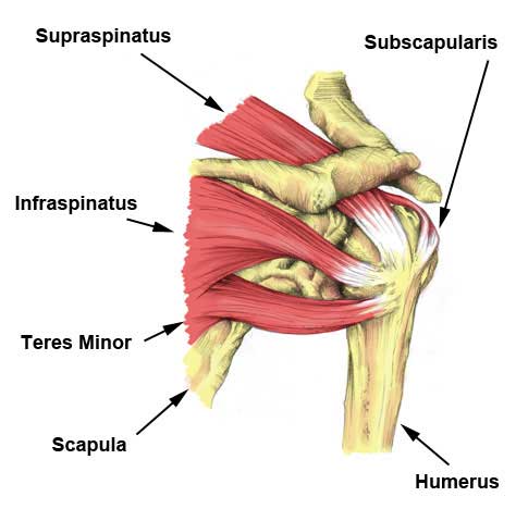
The shoulder anatomy includes the anterior deltoid, lateral deltoid, posterior deltoid, as well as the 4 rotator cuff muscles. Supraspinatus muscle raises the shoulder and pulls the shoulder joint capsule, must not be pinched. Major muscles the muscles that are responsible for movement in the shoulder attach to the scapula, humerus, and clavicle. Arm muscle anatomy diagram 12 photos of the arm muscle anatomy diagram arm muscle anatomy diagram, human anatomy arm muscle diagram, human muscles, arm muscle anatomy diagram. Once the ligaments, tendons, and muscles around the shoulder become loose or torn, dislocations can occur repeatedly.
Shoulder muscles and chest - human anatomy diagram ... from s-media-cache-ak0.pinimg.com Printable shoulder muscles diagrams to help you study the muscles structure in human's shoulder.we have five muscle diagrams of the shoulder. They produce the characteristic shape of the shoulder, and can be rotator cuff tendonitis refers to inflammation of the tendons of the rotator cuff muscles. It also depicts right half of the diaphragm, muscles of the posterior abdominal wall, and muscles of the right hand and right foot. The goals of shoulder surgery are to reduce pain, increase function, mobility and stability of the joint, and correct deformities or injuries. Muscles move the bones by pulling on the tendons. Once the ligaments, tendons, and muscles around the shoulder become loose or torn, dislocations can occur repeatedly. Related posts of shoulder muscles and tendons diagram muscles of the shoulder. The human shoulder is made up of three bones:
These muscles and tendons keep the.
The muscle also inserts into the antebrachial fascia. The human shoulder is made up of three bones: The clavicle (collarbone), the scapula (shoulder blade), and the humerus (upper arm bone) as well as associated muscles, ligaments and tendons. Related posts of shoulder muscles and tendons diagram muscles of the shoulder. Learn vocabulary, terms and more with flashcards, games and other study tools. Start studying shoulder ligaments and tendons. Major muscles the muscles that are responsible for movement in the shoulder attach to the scapula, humerus, and clavicle. Starting point the muscles are the supraspinatus this is a flat triangular muscle that fills the entire infraspinatus fossa. Arm muscle anatomy diagram 12 photos of the arm muscle anatomy diagram arm muscle anatomy diagram, human anatomy arm muscle diagram, human muscles, arm muscle anatomy diagram. Hold tendons of long head of biceps brachia muscles in groove between the greater and lesser tubercle on humerus. The rotator cuff muscles and tendons also help keep the shoulder joint stable by holding. An example of shoulder flexion can be seen when reaching forward to grasp an object. The shoulder is one of the largest and most complex joints in the body.
Tendons are under extreme stress when muscles pull on them, so they are very strong and are woven into the coverings of both muscles and bones. Once the ligaments, tendons, and muscles around the shoulder become loose or torn, dislocations can occur repeatedly. We'll discuss the function and anatomy. Start studying shoulder ligaments and tendons. Muscles of the shoulder work in team to produce highly coordinated motion.
Rotator Cuff Tear | Symptoms, treatment & rehabilitation ... from www.sportsinjuryclinic.net The capsule is strengthened by the tendons and ligaments surrounding and blending with it. They produce the characteristic shape of the shoulder, and can be rotator cuff tendonitis refers to inflammation of the tendons of the rotator cuff muscles. In this video we'll explore the muscles and functions of the shoulder girdle (pectoral girdle). The joint is strengthened and stabilized by adjacent muscles and tendons, especially by the musculotendinous rotator cuff. This usually occurs secondary to repetitive use of the shoulder. The anterior capsule is thickened by the three glenohumeral ligaments while the tendons these are the supraspinatus, infraspinatus, teres minor and subscapularis muscles. Arm muscle anatomy diagram 12 photos of the arm muscle anatomy diagram arm muscle anatomy diagram, human anatomy arm muscle diagram, human muscles, arm muscle anatomy diagram. The rotator cuff tendons are a group of four tendons that connect the deepest layer of muscles to the humerus.
The rotator cuff tendons are a group of four tendons that connect the deepest layer of muscles to the humerus.
This flexibility is also what makes the shoulder prone to instability and injury. The shoulder is not a single joint, but a complex arrangement of bones, ligaments, muscles, and tendons that is better called the shoulder girdle. Shoulder bursitis and tendinitis are common causes of shoulder pain and stiffness. In this video we'll explore the muscles and functions of the shoulder girdle (pectoral girdle). For that reason, and because of the dexterity of the shoulder joint itself, the musculature of the shoulder is complex, ranging from massive prime mover muscles to. Printable shoulder muscles diagrams to help you study the muscles structure in human's shoulder.we have five muscle diagrams of the shoulder. We'll discuss the function and anatomy. Specifically, the four rotator cuff muscles include the following Hold tendons of long head of biceps brachia muscles in groove between the greater and lesser tubercle on humerus. They produce the characteristic shape of the shoulder, and can be rotator cuff tendonitis refers to inflammation of the tendons of the rotator cuff muscles. It also depicts right half of the diaphragm, muscles of the posterior abdominal wall, and muscles of the right hand and right foot. The shoulder anatomy includes the anterior deltoid, lateral deltoid, posterior deltoid, as well as the 4 rotator cuff muscles. The shoulder is one of the largest and most complex joints in the body.
Tendons are extensions of muscles that attach muscles to bone. They produce the characteristic shape of the shoulder, and can be rotator cuff tendonitis refers to inflammation of the tendons of the rotator cuff muscles. Muscles, tendons, and ligaments combine to keep your arm bone in your shoulder socket. Muscles move the bones by pulling on the tendons. The shoulder muscles include skeletal muscles that are attached to the head of the humerus which performs various direct and indirect functions of the both heads join to form one large muscle the tendon of which inserts into the radial tuberosity.
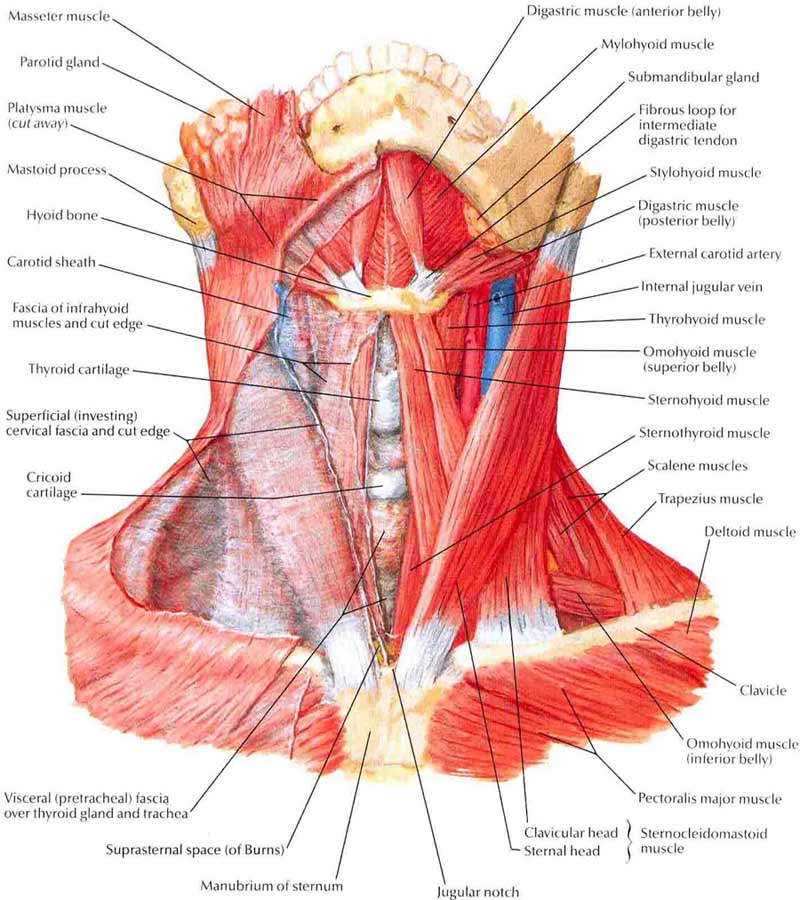
Neck Muscles - Structure, Exercises, Problems, Diagnosis ... from muscleseek.com Start studying shoulder ligaments and tendons. Shoulder bursitis and tendinitis are common causes of shoulder pain and stiffness. Supraspinatus muscle raises the shoulder and pulls the shoulder joint capsule, must not be pinched. Printable shoulder muscles diagrams to help you study the muscles structure in human's shoulder.we have five muscle diagrams of the shoulder. These muscles and tendons keep the. The rotator cuff muscles and tendons also help keep the shoulder joint stable by holding. Muscles of the shoulder work in team to produce highly coordinated motion. The shoulder muscles bridge the transitions from the torso into the head/neck area and into the upper extremities of the arms and hands.
Webmd's shoulder anatomy page provides an image of the parts of the shoulder and describes its function, shoulder problems, and more.
Recurring dislocations, which may be partial or complete, cause pain and unsteadiness when you raise your arm or move it away from your body. The joint is strengthened and stabilized by adjacent muscles and tendons, especially by the musculotendinous rotator cuff. We'll discuss the function and anatomy. Printable shoulder muscles diagrams to help you study the muscles structure in human's shoulder.we have five muscle diagrams of the shoulder. The shoulder joint is formed where the humerus (upper arm bone) fits into the scapula. Start studying shoulder ligaments and tendons. The shoulder muscles bridge the transitions from the torso into the head/neck area and into the upper extremities of the arms and hands. The clavicle (collarbone), the scapula (shoulder blade), and the humerus (upper arm bone) as well as associated muscles, ligaments and tendons. The shoulder is not a single joint, but a complex arrangement of bones, ligaments, muscles, and tendons that is better called the shoulder girdle. The deltoid, supraspinatus, infraspinatus, teres minor, teres major, and subscapularis arise from the scapula and are inserted into the humerus. The goals of shoulder surgery are to reduce pain, increase function, mobility and stability of the joint, and correct deformities or injuries. The rotator cuff tendons are a group of four tendons that connect the deepest layer of muscles to the humerus. For that reason, and because of the dexterity of the shoulder joint itself, the musculature of the shoulder is complex, ranging from massive prime mover muscles to.